Latest proton therapy study shows hypofractionated proton therapy as safe and effective for patients with low-risk prostate cancer

A study published in August 2019 in the International Journal of Particle Therapy by Dr. Slater and his team, highlights the results of their latest phase I/II hypofractionated proton therapy study at Loma Linda University Hospital.

Prostate cancer is the most commonly diagnosed cancer in men, and many of these patients have low-risk, early disease. Prostate cancer at these stages remains highly treatable with local control rates over 90% and very low rates of late morbidity commonly reported for a variety of treatment modalities. The focus then turns to the avoidance of unnecessary negative treatment-related side effects that can occur, particularly through the use of conventional treatments such as surgery and x-ray (conventional) radiotherapy.

Proton radiation therapy has demonstrated itself to be an excellent option for low-risk prostate cancer as it delivers high control rates with very little toxicity. Proton beam thereby enhances the physician’s opportunity to minimise risks for the patient.

Hypofractionation is the process of delivering higher doses of radiation per fraction, but using fewer daily fractions. Doctors and physicists at Loma Linda University have successfully used hypofractionated proton therapy for several diseases, including cancers of the breast, lung, and liver. In each instance, control and survival rates have been maintained and unwelcome side effects have not increased. This experience prompted the medical team at Loma Linda to investigate hypofractionation for prostate cancer.

The purpose of the study was to determine whether a hypofractionated proton radiotherapy regimen can control early-stage prostate cancer while maintaining low rates of side effects similar to results obtained using standard-fraction proton radiotherapy.

A cohort of 146 patients with low-risk prostate cancer (Gleason score 7, prostate-specific antigen 10, tumor stage of T1–T2a) received 20 fractions of proton therapy (3.0 Gy per fraction over 4 weeks). Patients were evaluated at least weekly during treatment, at which time documentation of treatment tolerance and acute reactions was obtained. Follow-up visits were conducted every 3 months for the first 1 years, every 6 months for the next 3 years, then annually. Follow-up visits consisted of history and physical examination, PSA measurements, and evaluation of toxicity.

The 3-year biochemical progression-free survival rate was 99.3%, and the 5-year biochemical progression-free survival was 97.9%.

In conclusion, this study showed that hypofractionated proton therapy (60 Gy in 20 fractions) was safe and effective for patients with low-risk prostate cancer. A prospective multi-institutional randomised study is currently being conducted to confirm these results.

Sources:

Kil WJ, Nichols RC Jr, Hoppe BS, Morris CG, Marcus RB Jr, Mendenhall W, Mendenhall NP, Li Z, Costa JA, Williams CR, Henderson RH. Hypofractionated passively scattered proton radiotherapy for low- and intermediate-risk prostate cancer is not associated with post-treatment testosterone suppression. Acta Oncol. 2013;52:492–7

Mendenhall NP, Hoppe BS, Nichols RC, Mendenhall WM, Morris CG, Li Z, Su Z, Williams CR, Costa J, Henderson RH. Five-year outcomes from 3 prospective trials of image-guided proton therapy for prostate cancer. Int J Radiat Oncol Biol Phys. 2014;88:596–602

Mendenhall NP, Li Z, Hoppe BS, Marcus RB Jr, Mendenhall WM, Nichols RC, Morris CG, Williams CR, Costa J, Henderson R. Early outcomes from three prospective trials of image-guided proton therapy for prostate cancer. Int J Radiat Oncol Biol Phys. 2012;82:213–21

Shipley WU, Verhey LJ, Munzenrider JE, Suit HD, Urie MM, McManus PL, Young RH, Shipley JW, Zietman AL, Biggs PJ, Heney NM, Goitein M. Advanced prostate cancer: the results of a randomized comparative trial of high dose irradiation boosting with conformal protons compared with conventional dose irradiation using photons alone. Int J Radiat Oncol Biol Phys. 1995;32:3–12

Slater JD, Rossi CJ Jr, Yonemoto LT, Bush DA, Jabola BR, Levy RP, Grove RI, Preston W, Slater JM. Proton therapy for prostate cancer: the initial Loma Linda University experience. Int J Radiat Oncol Biol Phys. 2004;59:348–52

Slater JD, Rossi CJ Jr, Yonemoto LT, Reyes-Molyneux NJ, Bush DA, Antoine JE, Miller DW, Teichman SL, Slater JM. Conformal proton therapy for early-stage prostate cancer. Urology. 1999;53:978–84

Slater JD, Yonemoto LT, Rossi CJ Jr, Reyes-Molyneux NJ, Bush DA, Antoine JE, Loredo LN, Schulte RW, Teichman SL, Slater JM. Conformal proton therapy for prostate carcinoma. Int J Radiat Oncol Biol Phys. 1998;42:299–304

Slater JM, Slater JD, Kang JI, et al. Hypofractionated Proton Therapy in Early Prostate Cancer: Results of a Phase I/II Trial at Loma Linda University. Int J Part Ther. 2019;6(1):1–9. doi:10.14338/IJPT-19-00057

Zietman AL, Bae K, Slater JD, Shipley WU, Efstathiou JA, Coen JJ, Bush DA, Lunt M, Spiegel DY, Skowronski R, Jabola BR, Rossi CJ. Randomized trial comparing conventional-dose with high-dose conformal radiation therapy in early-stage adenocarcinoma of the prostate: long-term results from Proton Radiation Oncology Group/American College of Radiology 95-09. J Clin Oncol. 2010;28:1106–11

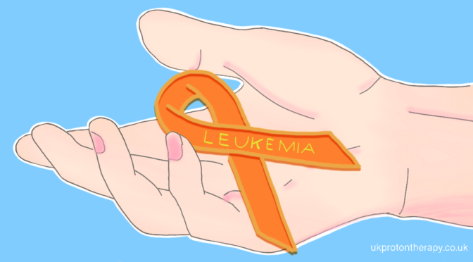
Proton Therapy for Central Nervous System Leukaemia

Interview with Dr Kateřina Dědečková on the possibilities of treatment of hematologic diseases with proton radiotherapy.

According to the latest recommendations of the International Lymphoma Radiation Oncology Group (ILROG), radiotherapy is also suitable for some patients diagnosed with leukaemia. Which patients specifically?

ILROG is very active in raising awareness on the benefits of radiation in the treatment of hematological malignancies, i.e. blood tumours. Over the past few years, ILROG has issued recommendations for the use of radiation in most haematologic cancers such as lymphomas, leukemias, myelosarcomas, myeloma, and others. For leukemias, in particular patients with central nervous system (CNS) involvement, it is now recommended to use radiation more frequently and to a greater extent.

Why is it now advisable to irradiate the central nervous system (CNS) also in patients with leukemia?

The CNS (brain and spinal cord) is separated from the bloodstream by a safety barrier, called blood–brain barrier (BBB), which prevents materials from the blood from entering the brain. Thus, some molecules, including drugs, reach the CNS with limited or no effect. For this reason, in some cases, the CNS may be a source of disease recurrence because part of the cells escapes the effect of chemotherapy or biological therapy, leading to a re-spread of cancer cells in the body.

What benefits do patients have from using proton radiotherapy?

Due to the limited possibilities of systemic treatment, irradiation of the entire CNS area is advantageous. This consists in irradiation of the brain, spinal cord, spaces where cerebrospinal fluid (CSF) circulates, craniospinal axis. The aim is also to get to the hard-to-reach leukaemia cells, as well as those that are no longer sensitive to chemotherapy or biological therapy.

Patients who benefit the most from proton radiotherapy are the ones who, despite intensive systemic treatment, have

  • a positive finding of leukemic cells in the cerebrospinal fluid;
  • a positive cerebrospinal fluid at the time of diagnosis;
  • leukemic lesions in the brain or spinal cord.

Also, patients who are at risk of involvement of the central nervous system future.

Therefore, according to the new recommendations, these patients should preferably be irradiated in the whole area of ​​the craniospinal axis.

Is radiotherapy commonly indicated in these patients or is it a new recommendation?

Previously, because of the high toxicity, it was preferable to irradiate only the skull area. However, thanks to the use of proton radiotherapy instead of classical radiation, it is possible to reduce the adverse effects of treatment that previously impaired quality of life, such as nausea, vomiting, fatigue, swallowing pain, and aphthae in the oral cavity. Therefore, radiotherapy has not been commonly used in these patients and, if so, less extensive exposure has been used, mainly because of concerns about toxicity associated with older exposure techniques, as I have mentioned. Irradiation of the CNS as a risk area has been found to improve the outlook for cure in risk patients. According to the new ILROG recommendation, groups of patients have already been identified who will benefit from the inclusion of craniospinal axis irradiation in their treatment plan.

How does the treatment work?

The patient is placed in supine position with their arms along the body, while the head is fixed with a special thermoplastic mask. A CT scan is done over the entire irradiation range, i.e. the entire head and spine to the coccyx. These CT images then show the areas that are the target of radiation and also the areas that we want to protect from radiation, called organs at risk (OAR). Then, the doctors and physicists collaborate to carefully calculate the irradiation plan. This plan is then checked multiple times, as well as directly in the gantry to confirm its accuracy. Then, the actual treatment begins. Leukaemia is typically treated in 9 to 12 fractions (sessions), applied to the entire craniospinal axis. In some patients, we also irradiate apparent tumor deposits, up to a total of 15-18 fractions. Irradiation takes place every weekday and may be associated with mild swallowing difficulties, fatigue and a decrease in the number of blood cells.

Is there a difference if a patient is irradiated with photon or proton techniques?

Yes, the difference is that when using proton RT, the patient is less exposed to radiation. There is less irradiation to the organs in front of the tumour, minimal irradiation to lungs, heart, oesophagus, intestinal loops, kidneys, liver, bladder, which significantly reduces the possibility of late toxicity of these organs, and the patient also better tolerates irradiation of craniospinal axis.

Do you have any recent experience with this form of treatment? If so, how did the patients tolerate the treatment?

At the Proton Therapy Center in Prague we already have experience with this irradiation technique for hematological malignancies. In some of these patients, RT was the last treatment option and even here we managed to successfully destroy CNS tumor cells. From our point of view this is a very promising method of treatment, but a longer follow-up of patients and evaluation of a larger group of patients will be required.

Dr Kateřina Dědečková

In 1998-2001 Dr Dědečková worked at the radiotherapy department of the District Hospital in Jičín. Then, from 2001 to 20017, she worked at the Institute of Radiation Oncology of the University Hospital Na Bulovce. In 2009 Dr Dědečková gained specialised competence in the field of radiation oncology. Since 2012, she has been working as a radiation oncologist at the Proton Therapy Center in Prague. Since 2019, Dr Dědečková also leads the interdisciplinary Center of Excellence for Proton Radiotherapy of Malignant Lymphomas at the Proton Center in Prague.

Dr Dědečková specializes in radiation treatment of malignant lymphomas and other hematological tumours, urological tumours and head and neck tumours. She has participated in international clinical trials with independent quality control of radiotherapy (GHSG, ESTRO Equal, QARC) in the treatment of malignant lymphomas and head and neck tumours. Dr Dědečková is a member of the Council of the International Lymphoma Radiation Oncology Group (ILROG) and a member of the Lymphoma Sub-Committee of the Particle Therapy Co-Operative Group (PTCOG). Dr Dědečková publishes professional medical press and lectures at congresses, both domestically and internationally. Dr Dědečková is co-author of the “Diagnostic and Treatment Guidelines in Patients with Malignant Lymphoma” of the Lymphoma Cooperative Group (chapter on lymphoma radiotherapy). Dr Dědečková deals with new techniques of lymphoma radiotherapy, such as proton radiotherapy of mediastinal lymphomas using the pencil beam scanning technique in maximum inhalation.

New study shows proton therapy more effective than conventional radiotherapy in the treatment of intrahepatic cholangiocarcinoma (ICC)

In a recent study by the Massachusetts General Hospital Department of Radiation Oncology, the use of proton therapy for intrahepatic cholangiocarcinoma (ICC) was shown to more effectively control the cancer and improve the chances of survival – particularly in comparison to conventional (photon) radiotherapy.

Cholangiocarcinoma is a cancer that develops in the cells within the bile ducts; both inside and outside the liver. The terms cholangiocarinoma and bile duct cancer are often used to refer to the same condition. This condition occurs slightly more often in males than females and usually affects people who are between 50-70 years old. Signs and symptoms of intrahepatic cholangiocarcinoma include jaundice, abdominal pain, fever, weight loss, weakness and itching. Treatment options may include surgery (when possible) to remove the bile duct and parts of the liver, chemotherapy and radiation.

In certain cases cholangiocarcinoma is an unresectable form of cancer. Unresectable cancer is defined as a cancer or tumour that cannot be removed completely through surgery. In these cases, radiotherapy and chemotherapy offer the best chances of survival.

The aim of this study was to evaluate outcomes for patients with unresectable intrahepatic cholangiocarcinoma (ICC) treated with hypofractionated proton or photon radiation therapy.

66 patients with unresectable intrahepatic cholangiocarcinoma were treated with hypofractionated proton (32 patients) or photon (34 patients) radiation therapy. Median radiotherapy (RT) dose was 58.05 Gy, all delivered in 15 daily fractions. On multivariate analysis for overall survival (OS), compared with photon RT, there was a trend towards improved survival with proton RT (HR 0.50; p = 0.05).

Median follow-up times from diagnosis and RT start were 21 months and 14 months, respectively. In total, five patients (7.6%) developed local failure. The 2-year outcomes were 84% local control (LC) and 58% OS. Among the 51 patients treated with definitive intent, the 2-year LC rate was 93% and the OS rate was 62%.

The study concluded that hypofractionated radiation therapy yields high rates of local control and is an effective modality to optimize biliary control for unresectable/locally recurrent ICC.

At the ESMO World Congress on Gastrointestinal Cancer 2019, it was identified that high dose radiotherapy in unresectable ICC should be considered as a viable treatment option, in combination with systemic therapy.

This study adds to the growing body of evidence suggesting proton beam therapy as a safe and effective treatment for patients with unresectable ICC. It is proposed therefore, that – pending further research – proton therapy be utilised as a curative treatment for ICC.

Sources:

Hong TS, Wo JY, Yeap BY, Ben-Josef E, McDonnell EI, Blaszkowsky LS, Kwak EL, Allen JN, Clark JW, Goyal L, et al. Multi-institutional phase II study of high-dose Hypofractionated proton beam therapy in patients with localized, Unresectable hepatocellular carcinoma and intrahepatic Cholangiocarcinoma. J Clin Oncol. 2016;34(5):460–8

Ohkawa A, Mizumoto M, Ishikawa H, Abei M, Fukuda K, Hashimoto T, Sakae T, Tsuboi K, Okumura T, Sakurai H. Proton beam therapy for unresectable intrahepatic cholangiocarcinoma. J Gastroenterol Hepatol. 2015;30(5):957–63

Shimizu, S., Okumura, T., Oshiro, Y. et al. Clinical outcomes of previously untreated patients with unresectable intrahepatic cholangiocarcinoma following proton beam therapy. Radiat Oncol 14, 241 (2019)

Smart AC, Goyal L, Horick N, Petkovska N, Zhu AX, Ferrone CR, Tanabe KK, Allen JN, Drapek LC, Qadan M, Murphy JE, Eyler CE, Ryan DP, Hong TS, Wo JY. Hypofractionated Radiation Therapy for Unresectable/Locally Recurrent Intrahepatic Cholangiocarcinoma. Ann Surg Oncol. 2019 Dec 23. doi: 10.1245/s10434-019-08142-9 [Epub ahead of print]

Ustundag Y, Bayraktar Y. Cholangiocarcinoma: a compact review of the literature. World J Gastroenterol. 2008;14(42):6458–66